
"It's Just Period Pain — It Will Pass" Is a Dangerous Assumption
When period pain strikes, the advice is almost always the same: put a hot-water bottle on your belly, drink brown sugar water, take a painkiller, and wait. "Period pain is normal," goes the reassurance. "It will get better after you have a baby."
For many women, mild cramping is indeed ordinary. But when the pain follows a predictable pattern, returns month after month, and steadily intensifies, it may be the visible tip of something far more complex — endometriosis.
For Ms. J, a Korean professional working in China, that "it will pass" mindset nearly cost her far more than a few uncomfortable days. Months of ignored pain ended in a suspected ruptured chocolate cyst with severe pelvic adhesions. What followed was a race to protect her ovaries — and her future fertility.
A Missed Warning: Ms. J's Story
Ms. J had never suffered from period pain before. In June 2026, the pain arrived suddenly. She assumed it was temporary and endured it. But the pattern began to repeat: two to three days before every period, the cramps returned.
In August, while visiting Korea, the pain intensified. A local gynecologic ultrasound raised the possibility of ovarian endometriosis, and her tumor markers were elevated across the board. Her doctors suspected a ruptured chocolate cyst. Pain-relief injections eased the symptoms, but the underlying problem remained.
Because she had long worked in China and was more familiar with the Chinese healthcare system, Ms. J chose to seek treatment there. In early September, she secured an appointment with Professor Sun Dawei, a leading gynecologic surgeon at AMCARE Beijing.
MRI Revealed More Than Expected
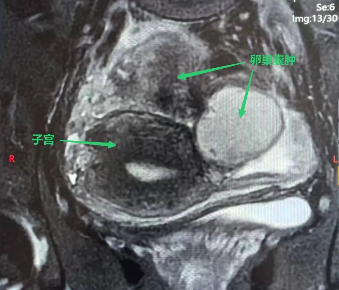
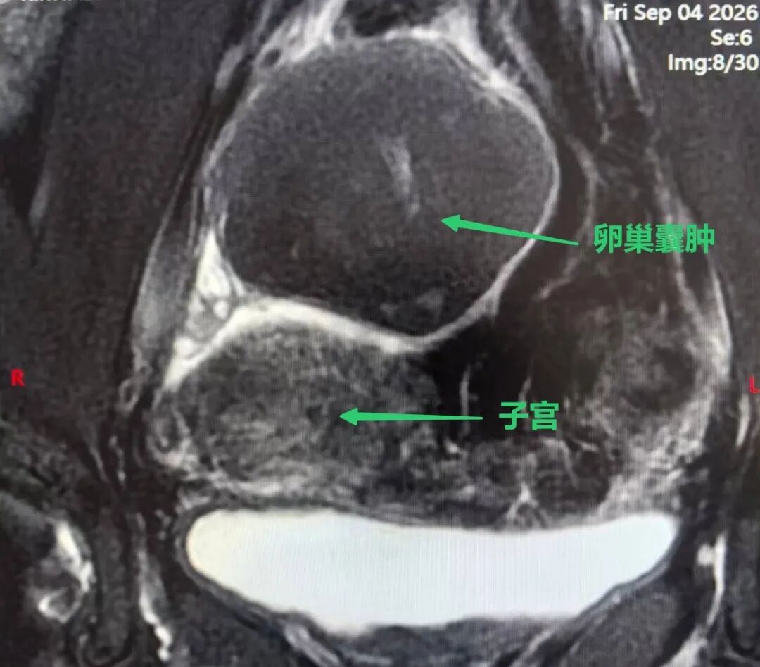
Professor Sun arranged a pelvic MRI. The findings were more complicated than the initial ultrasound had suggested:
- Both ovaries were enlarged, with nodular lesions and hemorrhage — consistent with bilateral endometriomas (chocolate cysts).
- Focal endometriosis with possible hemorrhage was seen on the posterior wall of the uterus.
- A small uterine fibroid and pelvic fluid were also present.
Combining the imaging with her history, Professor Sun concluded that the cyst had already ruptured. "Chocolate"-like fluid had spilled into the abdominal cavity and was triggering widespread pelvic adhesions and pain. Surgery needed to be scheduled promptly.
Inside the "One-Inch" Battlefield
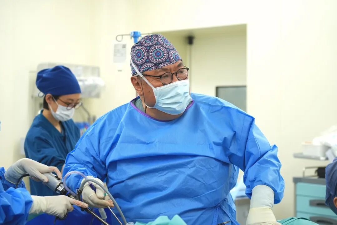
In the operating room, Professor Sun introduced a single-port laparoscopic camera through a 2 cm incision at the umbilicus. What appeared on the screen was not a clean pelvis — it was, in his words, "a complete mess."
- The omentum, small bowel, uterine surface and both adnexa were bound together by extensive membranous adhesions, wrapping the uterus, ovaries and pelvic floor.
- Patches of brown, viscous fluid were scattered across the pelvic wall; the colon was stuck to the left pelvic wall.
- The right ovary carried a cyst of about 6 cm, densely adherent to the posterior uterine wall and pelvic floor — it could not even be lifted.
- The left ovary was cystically enlarged to about 3 cm, densely adherent to the left posterior wall of the uterus.
Ms. J had not yet had children. For the surgical team, preserving ovarian function was the highest principle — and that made an already difficult operation even more demanding.
Adhesiolysis First, Then Cystectomy
Free the adhesions before removing the cysts. Because the omentum, bowel, uterus and both adnexa were extensively adherent, the team first had to separate the surrounding organs with blunt and sharp dissection before the ovaries could be exposed and mobilized. This stage tests the surgeon's patience: a moment of carelessness can injure the bowel or major vessels.
Single-port access raises the difficulty another notch. Every instrument must enter through the same 2 cm umbilical opening. The resulting "chopstick effect" — instruments crowding and interfering with one another — places high demands on surgical skill and spatial imagination.
At every step, the team was leaving room for fertility:
Minimize Thermal Injury
Careful electrocoagulation limited heat spread to the ovarian tissue, protecting the healthy cortex that must remain functional after surgery.
Prevent Re-Adhesion
An anti-adhesion barrier was placed to reduce the risk of the fallopian tubes and ovaries scarring together again after the procedure.
Preserve Cosmesis
The single umbilical incision hides the scar within the natural fold of the navel, leaving the abdomen essentially unmarked.
Speed Recovery
An ERAS (enhanced recovery after surgery) protocol reduced bed rest and immobility — both of which can contribute to new adhesions.
The Outcome: 51 Minutes, About 5 ml of Blood Loss
In the end, the cyst walls on both ovaries were completely removed and extracted through the umbilical incision. The operation took 51 minutes, and total blood loss was only about 5 ml.
Procedure Snapshot
- Approach: Single-port laparoscopy via a ~2 cm umbilical incision
- Findings: Bilateral endometriomas (6 cm right, 3 cm left) with severe pelvic adhesions
- Procedure: Adhesiolysis and bilateral ovarian cystectomy, ovarian tissue preserved
- Duration: 51 minutes
- Estimated blood loss: About 5 ml
Surgery Is Only the First Step
"Completing the operation is only the beginning," Professor Sun noted. "Without long-term management, endometriomas have a high recurrence rate." To reduce that risk, he designed a long-term medical management plan for Ms. J, with regular follow-up pelvic imaging and monitoring of liver and kidney function.
Period Pain Is a Disease — Not a Virtue
Reflecting on the case, Dr. Li Shujuan, Director of Gynecology at AMCARE, emphasized that the frightening part was not the chocolate cyst itself, but the mindset of "just endure it and it will pass."
About one in three women with endometriosis has no obvious symptoms. Many others mistake pathological period pain for a normal physiological response — and wait years before seeking help. An endometrioma can take years to grow to the size at which it ruptures. Regular gynecologic check-ups, including pelvic ultrasound, are the key to early detection and early intervention, buying precious time before the disease progresses.

In the face of endometriosis, endurance is not a virtue — it is a blade suspended over a woman's health. The longer it is ignored, the greater the physical and emotional toll.
What Is a Chocolate Cyst?
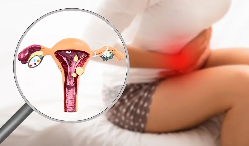
A chocolate cyst is a common form of endometriosis. Normally, the endometrium lines the inside of the uterine cavity. Each month, under the influence of hormones, it thickens, secretes, and is shed during menstruation.
When endometrial tissue grows outside the uterine cavity and forms a lesion, the condition is called endometriosis. When that tissue settles on the ovary, it still responds to the cyclical rise and fall of estrogen and progesterone. Every menstrual cycle brings another bleed — but the blood has nowhere to go. It accumulates, forming an old blood-filled sac whose contents resemble chocolate. Hence the name chocolate cyst.
Three Warning Signs That Warrant a Doctor's Visit
If any one of the following applies, seek a gynecologic evaluation promptly:
- Progressively worsening pain — each period is more painful than the last.
- Painkillers stop working, or increasingly large doses are needed for relief.
- Pain interferes with daily life, or is accompanied by infertility or pelvic pain outside menstruation.
"Endometriosis does not disappear because we ignore it. The earlier it is recognized, the more options — and the more ovarian reserve — a woman still has."
— Dr. Li Shujuan, Director of Gynecology, AMCARE
Reviewed by
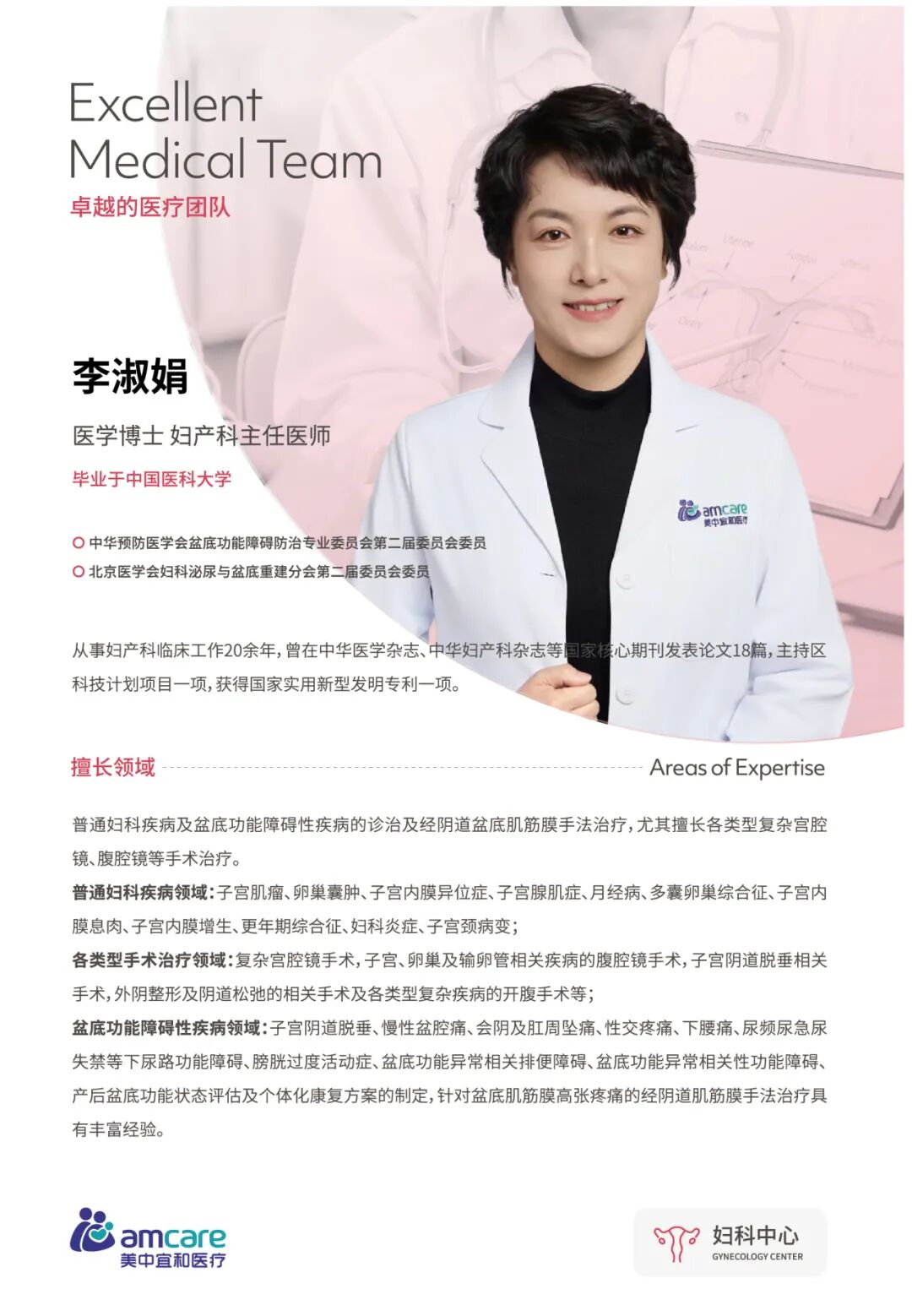
Dr. Li specializes in general gynecologic and pelvic floor disorders and in complex laparoscopic and hysteroscopic surgery, including ovarian cysts, endometriosis, uterine fibroids, adenomyosis, menstrual disorders, polycystic ovary syndrome, endometrial polyps, and cervical lesions.
Need Help With Severe Period Pain or a Suspected Ovarian Cyst?
AMCARE Beijing's gynecology and international patient teams provide ultrasound review, second opinions, fertility-preserving minimally invasive surgery, and coordinated care for women traveling to China for treatment.