
Why Pregnancy Ultrasound Is More Than "One and Done"
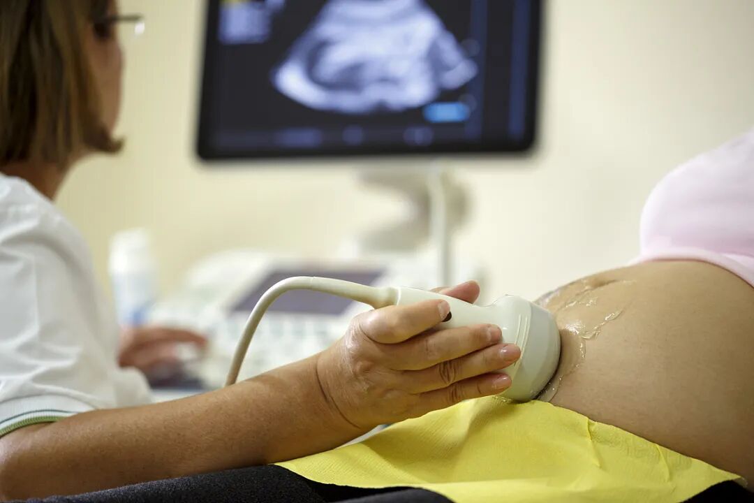
Ultrasound is the most widely used imaging tool in pregnancy. It uses harmless sound waves to create real-time pictures of the baby, placenta, amniotic fluid and uterus. For expectant parents, it is also one of the most anticipated moments of prenatal care.
However, pregnancy ultrasound is not a single event. It is a series of timed examinations spread across the three trimesters, each designed to answer different clinical questions. Two of the most important — and most misunderstood — are NT screening in the first trimester and the mid-pregnancy anomaly scan. Many families also ask whether a glamorous 4D video can replace the anomaly scan. The short answer is: no.
First-Trimester Ultrasound
6–8 Weeks: Confirming Pregnancy and Ruling Out Ectopic Pregnancy
The first ultrasound is usually performed at 6–9 weeks. Its main goals are to:
- Confirm the pregnancy is inside the uterus and rule out ectopic pregnancy.
- For mothers with a prior cesarean section, rule out cesarean scar pregnancy.
- Check gestational sac location and size, look for fetal pole and heartbeat, determine single or multiple pregnancy, and confirm gestational age.
Embryos are usually visible around 45 days, and cardiac activity is typically detectable at 6–8 weeks, so scanning too early can miss reassuring findings.
11–13 Weeks + 6 Days: NT (Nuchal Translucency) Screening
NT screening is one of the most important first-trimester tests. It measures the thickness of fluid at the back of the fetal neck and combines the result with maternal blood screening to estimate the risk of chromosomal abnormalities such as Down syndrome.
The scan also allows the physician to:
- Count fetuses and confirm viability.
- Observe the fetal heartbeat and major structures such as the limbs, spine and cranium.
- Detect some early severe structural anomalies.
Important Timing Window
NT measurement is accurate only between 11 weeks + 0 days and 13 weeks + 6 days. After 14 weeks the fluid is normally reabsorbed, so the result is no longer reliable.
Second-Trimester Ultrasound
15–20 Weeks: Routine Mid-Pregnancy Ultrasound
During this window, the physician checks whether fetal growth matches gestational age, evaluates fetal movement, heart activity, placenta position and amniotic fluid volume, and integrates the results with maternal serum screening or non-invasive prenatal testing (NIPT).
20–24 Weeks + 6 Days: The Anomaly Scan ("Big Anomaly Scan")
This is the most comprehensive structural examination of the entire pregnancy. The optimal window is 22–24 weeks, when fetal organs are developed enough to visualize but there is still room to move.
The sonographer systematically examines the major fetal structures, including:
- Brain: ventricles and posterior fossa.
- Spine: alignment and closure.
- Face: lips and nose.
- Heart: four-chamber view and left/right ventricular outflow tracts.
- Chest and abdomen: diaphragm, abdominal wall, stomach bubble, kidneys, bladder.
- Limbs: long bones and hands/feet.
Biometric measurements (biparietal diameter, head circumference, abdominal circumference, femur length) are also taken to track growth. Placenta position, cord insertion and amniotic fluid are documented.
24–26 Weeks: Fetal Echocardiography
If the anomaly scan raises concerns about the heart, or if the mother has risk factors such as advanced maternal age, a family history of congenital heart disease, diabetes or infection during pregnancy, a dedicated fetal echocardiogram may be recommended for a detailed assessment of cardiac structure and function.
Passing the Anomaly Scan Does Not Mean 100% Certainty
Ultrasound cannot detect every abnormality. It is excellent for major structural defects, but small ventricular septal defects, minor digit anomalies, some cleft palate variants, and functional problems such as hearing, vision or intellectual disability usually cannot be identified before birth.
Third-Trimester Ultrasound
In the third trimester, the focus shifts from detecting structural anomalies to monitoring growth and well-being before delivery.
28–32 Weeks: Late-Pregnancy Growth Scan
This scan evaluates whether the fetus is growing appropriately for gestational age, checks fetal position (head-down or breech), reassesses placenta location (to rule out placenta previa), and measures amniotic fluid. Some centers also perform a late follow-up for anomalies that were not visible earlier.
36–37 Weeks: Term Assessment
Before delivery, the physician checks umbilical cord blood flow, amniotic fluid, placental maturity and fetal position, and estimates fetal weight. If the baby is breech, options to encourage a safer vaginal delivery can be discussed.
4D Ultrasound vs. Anomaly Scan: Don't Confuse the Two
Among all pregnancy ultrasounds, the anomaly scan carries the highest clinical value. Yet it is often confused with 4D ultrasound, which is popular for its clearer surface rendering and ability to produce short video clips and photos.
The two are fundamentally different:
- The anomaly scan is a diagnostic examination. Its core is two-dimensional (2D) grayscale imaging. The physician dynamically evaluates each structure and only briefly switches to 3D/4D when a specific coronal or surface view would improve diagnosis — for example, facial or limb defects where 3D rendering increases detection.
- 4D (live 3D) ultrasound is mainly a communication and keepsake tool. It creates realistic surface images that help parents bond with their baby. Short video clips and photos are for纪念 (memory-keeping), not diagnosis.
Real diagnostic decisions about organs, bones and internal structures are still made from the underlying 2D slices. In other words, 4D ultrasound may look more valuable visually, but the true clinical strength lies in the 2D anomaly scan.
If you would like a keepsake video, it can usually be recorded during the same appointment — after the formal anomaly scan is complete — as long as the fetal position and time allow.

Reviewed by
Medical Disclaimer
This article is for general information only and is not intended as medical advice, diagnosis or treatment. It does not replace a consultation with a qualified healthcare professional. Individual circumstances vary; if you are pregnant or planning pregnancy, please discuss your ultrasound schedule with your obstetrician.
Planning Prenatal Care in Beijing?
AMCARE Beijing's obstetrics and maternal-fetal medicine teams provide structured prenatal ultrasound screening, genetic counseling and international patient services for expectant families from overseas.