
The Package Is Not the Point
There is a reasonable assumption buried in the way most people buy a health check-up: that the more tests a package contains, the safer the person leaving with the report. The assumption survives because it is almost true. Almost — because the items in a package are chosen by a logic that has very little to do with the two events most people are actually afraid of.
Heart attack and stroke do not arrive as separate illnesses with separate causes. In the great majority of cases they are the same process in different plumbing: an atherosclerotic plaque that has been forming quietly for twenty or thirty years, in a coronary artery, in a carotid artery, in a vessel inside the skull. The plaque does not announce itself. It narrows the pipe, or it ruptures and a clot closes what was left, and the first symptom is the event itself.
That long silent interval is the entire argument for imaging. A vessel can be photographed while it is still merely abnormal — plaque present, wall thickened, flow beginning to change — and that photograph is worth something only if it changes what happens next. So the question worth asking about any check-up is not "how many items" but "which of these looks at the process that would kill me, and would the answer change anything".
What follows is an attempt to answer that question honestly, test by test, for the heart and the brain. It is also an explanation of why the most valuable items are frequently the cheapest — and the ones most often left out.
Why Packages Miss the Vessels
A check-up package is a commercial object before it is a clinical one. It is assembled to fit a price point and a morning's schedule, and the units it is assembled from are organs: liver, kidney, thyroid, lung, heart, uterus. Organ-based thinking produces a package that is thorough about organs and nearly blind to systems.
Atherosclerosis is a system. It does not respect the boundary between a cardiology department and a vascular one, and it usually appears in more than one place at once — a person with plaque in the carotid bulb very often has plaque in the femoral artery and in the coronary tree. The clinical name for this is polyvascular disease, and the practical consequence is that a single well-chosen ultrasound can tell you something about vessels you have not imaged.
There is a second reason for the gap, and it is logistical rather than intellectual. Ultrasound is cheap for the patient and expensive for the provider: it consumes fifteen to thirty minutes of a trained operator's hands, one patient at a time, and it cannot be batched. A contrast CT costs more per unit and runs a scanner on a schedule. A package optimised for throughput will therefore contain fewer ultrasounds than the evidence would recommend. None of this is a conspiracy; it is arithmetic, and it is the reason a patient who wants vascular screening usually has to ask for it by name.
Seven Tests for the Heart, and What Each One Is Looking At
These are the studies that belong to a credible cardiovascular screen. Not all of them are appropriate for everyone, and the sequence below is not a checklist to complete but a map of what each instrument can and cannot see.
1. Carotid ultrasound
The carotid arteries run up the neck and are the most accessible large vessels in the body, which is why they are used as a window onto the whole arterial system. The scan measures intima-media thickness, identifies plaques, describes their character — smooth and calcified or soft and lipid-rich, the latter being the more dangerous kind — and grades any narrowing. A carotid plaque is a marker of generalised atherosclerosis and is statistically linked to both stroke and coronary risk. It is painless, radiation-free, and takes about fifteen minutes.
2. Echocardiography
This is the structural and functional examination of the pump itself. It measures ejection fraction, chamber dimensions, wall thickness and valve function, and it shows whether a region of muscle is moving poorly because its blood supply has been reduced. It will not show the coronary arteries, and this is the most common misunderstanding about it: a normal echocardiogram means the pump is working, not that the pipes are clean. In hypertension it also documents the thickening of the left ventricle that years of high pressure produce.
3. Abdominal ultrasound of liver, gallbladder, pancreas and spleen
Not a cardiovascular test, but included for a reason that is easy to underestimate: a fatty liver is a metabolic marker that travels with insulin resistance, dyslipidaemia and hypertension, and the same visit is the natural moment to establish a baseline for organs that will be affected if vascular disease is found. Screening a system means knowing the metabolic ground it is growing in.
4. Urinary tract ultrasound
Structural screening of the kidneys, ureters and bladder. Its relevance here is not incidental. The kidneys are both a cause and a victim of vascular disease — renal artery narrowing drives blood pressure up, and long-standing hypertension damages the kidney — so a structural baseline, including kidney size and any obstruction, is part of the same picture. It also takes minutes and involves no contrast.
5. Ambulatory ECG (24-hour Holter)
A resting ECG is a ten-second photograph. Arrhythmias are often not there for those ten seconds. A Holter records the whole day and night and catches what a clinic visit systematically misses: paroxysmal atrial fibrillation, runs of premature beats, and transient episodes of ischaemia while the person is asleep, at work, or climbing stairs. Paroxysmal atrial fibrillation matters disproportionately here because it is a leading mechanism of embolic stroke in people whose resting ECG is entirely normal.
6. Ambulatory blood pressure monitoring
Twenty-four hours of blood pressure, taken every fifteen to thirty minutes by day and less often at night. It answers questions a single reading cannot: whether pressure falls at night the way it should, whether it surges in the early morning, whether the clinic reading was elevated because of the clinic. People whose pressure does not dip overnight — non-dippers — carry a higher cardiovascular risk at the same daytime number, and the pattern is invisible without the recording. It is also the fairest way to decide whether someone actually needs medication.
7. Coronary CT angiography
The only item on this list that images the coronary arteries directly. It produces a calcium score and, with contrast, a view of the lumen: whether plaque is present, how much, and whether any segment is significantly narrowed. Its strength is exclusion — a genuinely clean coronary tree in a symptomatic person is powerfully reassuring. Its costs are real and should be stated plainly: ionising radiation in the low single-digit millisievert range with modern protocols, an iodine-based contrast agent that requires reasonable kidney function and is unsuitable in pregnancy, a heart rate that may need to be slowed before the scan, and a finding of "moderate plaque" that can trigger further testing without changing management. It is a test to be chosen for a reason, not added for completeness.
Two More for the Brain
Stroke prevention has its own blind spot, and it sits inside the skull, where ultrasound cannot reach. Two magnetic resonance studies fill it, and neither uses ionising radiation.
Brain MRI without contrast shows what a CT scan tends to miss: small established infarcts, old micro-haemorrhages, white matter changes that accumulate with hypertension, small tumours, and the atrophy patterns of neurodegenerative disease. In a person with no symptoms it often produces findings that are best described as age-appropriate — a distinction that requires an experienced reader, because the difference between "expected for sixty" and "a warning" is the whole value of the study.
Brain MRA images the arteries themselves, without contrast in the time-of-flight technique most often used for screening. It detects narrowing, aneurysm and the anatomical variants that make some people more vulnerable to stroke, and it complements the carotid ultrasound: ultrasound sees the neck, MRA sees above it. A plaque at the carotid bifurcation and a stenosis of the middle cerebral artery are the same disease in two places, and only one of them shows up on a neck scan.
The Three That Are Almost Never Included
These three ultrasounds are inexpensive, radiation-free, and directly relevant to vascular risk. They are also the ones you will usually have to ask for, which is why clinicians who work in screening tend to call them by an unflattering name: the commonly omitted three.
Vertebral artery ultrasound. Two vertebral arteries run up the back of the neck and join to form the vessel that supplies the back of the brain — the territory responsible for balance, vision and coordination. Their first segment is reachable by ultrasound. The scan is recommended in men over forty with any of the familiar risk factors: abnormal lipids, smoking history, diabetes, hypertension. Vertebral arteries are frequently asymmetric by nature, so the value of the scan is not the presence of a difference but a documented baseline that a future study can be compared against.
Renal artery ultrasound. Renal artery stenosis is one of the few genuinely curable causes of hypertension, and it is worth excluding in anyone whose pressure is resistant, who is young at onset, or whose renal function drifted after starting an ACE inhibitor. Ultrasound is the reasonable first step because it is harmless and cheap, but its limitations should be understood: bowel gas and body habitus degrade the image, and a normal study does not fully exclude stenosis. When clinical suspicion is high, CT or MR angiography is the next move. The scan's other job is to look at the kidney itself — size, cortex, scarring — which is how the heart-kidney axis gets assessed in one sitting.
Bilateral lower-limb arterial ultrasound. Peripheral arterial disease is the most under-diagnosed manifestation of atherosclerosis, partly because it is silent for years and partly because the symptom people notice — cramping in the calf after walking a few hundred metres — is easily attributed to age or to the wrong department. Finding it matters beyond the legs: its presence roughly doubles the likelihood of coexistent coronary and cerebrovascular disease, and it changes a person's risk category. An ankle-brachial index takes two minutes and can precede the ultrasound; the scan then localises and grades what the index only hints at.
Put together, the three take well under an hour, require no contrast, expose no radiation, and cost less than a single contrast CT. That combination — high information, low risk, low price — is precisely the profile that package design tends to leave out, because none of those three qualities is what a package is selling.
How to Ask for It
For a reader planning this, four practical points decide whether a morning produces a usable answer or a folder of unrelated pictures.
Sequence matters. Blood pressure monitoring and Holter recording are fitted and returned, so they bookend the visit rather than sit inside it: the monitors go on at the start, the imaging happens during the day, the devices come off the next morning. A programme that understands this can fit a genuinely comprehensive screen into roughly thirty hours of elapsed time and one working day of presence.
Preparation is not optional. Abdominal and renal ultrasound require fasting; coronary CTA requires a recent creatinine value, a heart rate that can be controlled, and no metformin-related uncertainty in diabetic patients; MRI requires screening for metal, implants and claustrophobia. Arriving without these turns a scheduled slot into a rescheduled one.
Reports must be comparable. The value of a baseline is that the next study can be laid beside it. Ask for measurements, not adjectives — intima-media thickness in millimetres, stenosis as a percentage, calcium score as a number, ejection fraction as a percentage — and ask for the images themselves on a disc or in DICOM format. Patients who change country every few years discover the hard way that a report saying "mild plaque" cannot be compared to anything.
Have the conversation about what happens next. A screening test earns its place only when a positive result changes management: a statin started, a blood pressure regimen adjusted, an antiplatelet considered, a cardiology referral made, a follow-up interval set. If no one can describe that path before the scans are done, the scans are entertainment.
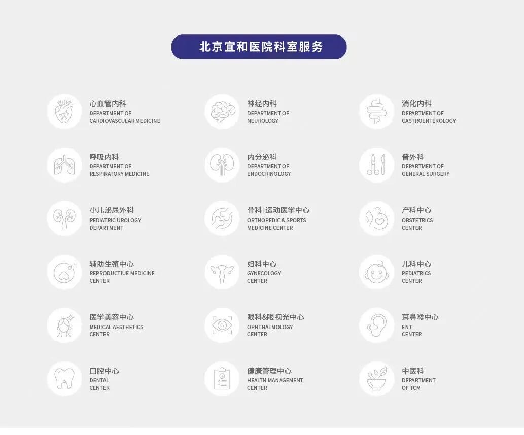
For international patients, the practical questions are usually about time and language: whether the whole set can be completed in one or two days, whether instructions and reports are issued in English, whether insurance documentation can be produced, and whether imaging can be taken away. AMCARE's health management center in Beijing runs these studies as a single booked pathway with English-language reports, and the international patient team can confirm the schedule and preparation requirements before travel.
Vessels Are Honest
The frightening thing about a heart attack or a stroke is not that it is sudden. It is that it is rarely sudden and almost always unannounced. Atherosclerosis gives notice for decades in a language nobody is listening for: a plaque forming, a wall thickening, a night-time pressure that no longer dips, a flow that has begun to slow.
The purpose of a well-chosen screen is to hear that language while it is still a whisper. Not to eliminate risk — nothing does — but to move the moment of discovery from the ambulance to a Tuesday morning in an ultrasound room, where the finding is a number on a report and the response can still be a tablet, a walk, and a follow-up in a year.
A check-up should not make life more complicated. It should make health more legible. The difference between those two outcomes is rarely the size of the package. It is whether the tests inside it were chosen to look at the thing you are actually afraid of.
This article describes general principles of cardiovascular and cerebrovascular screening and does not replace assessment by a treating physician. Which studies are appropriate depends on age, risk factors, previous results and contraindications; contrast studies in particular require individual review.