When Recovery Turns Into an Emergency
The six-hour surgery was finally over. A patient in her late twenties — we will call her Xiao Ai (a pseudonym) — had undergone bilateral adnexectomy, appendectomy, and lymph-node dissection for endometrial adenocarcinoma. She had already endured months of chemotherapy and radiotherapy. The operation was meant to remove the threat. Instead, four hours later, her heart delivered a new one.
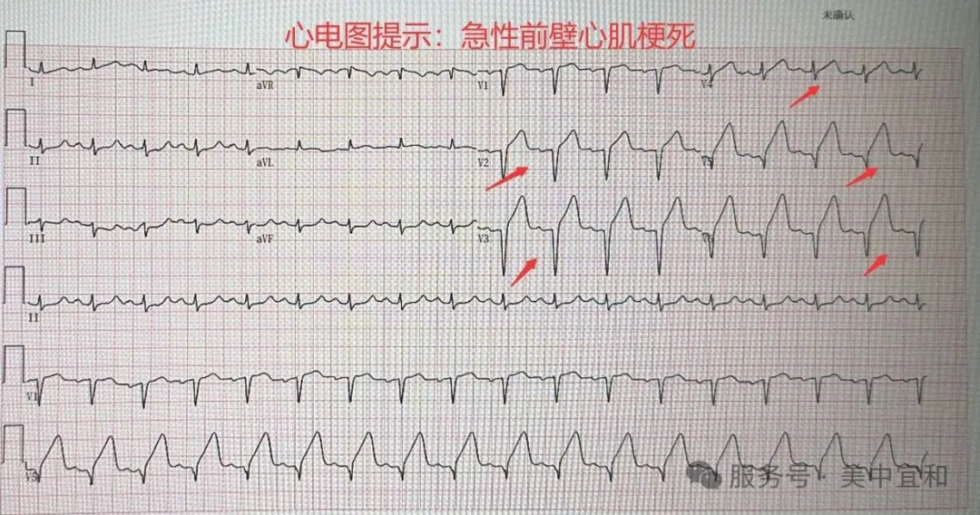
Back in the ward, Xiao Ai suddenly felt nauseous and vomited, with marked palpitations. An electrocardiogram shocked the team: ST-segment elevation in leads V1 through V6. An echocardiogram showed segmental wall-motion abnormality. Given her history of lower-limb venous thrombosis and the postoperative hypercoagulable state, the diagnosis pointed clearly to acute anterior myocardial infarction, likely thromboembolic.
She was young, with no hypertension, coronary disease, or classic crushing chest pain. The initial nausea had looked like ordinary postoperative gastroparesis. But myocardial ischemia does not wait for a convenient symptom. The local hospital arranged emergency transfer to AMCARE Beijing, and while the ambulance was en route, an MDT huddle was already assembling: cardiology, emergency medicine, gynecology, and medical affairs.
Three Paths, None of Them Safe
Acute MI after major abdominal surgery is rare and treacherous. The central conflict was stark: the clot in the coronary artery needed antithrombotic therapy, but the fresh surgical field needed hemostasis. The team mapped three options:
Option 1: Systemic Thrombolysis
Clot-dissolving drugs could reopen the vessel, but they require systemic anticoagulation. With extensive fresh surgical wounds inside the abdomen, uncontrolled postoperative bleeding was a terrifying risk.
Option 2: Emergency Percutaneous Coronary Intervention (PCI)
Coronary angiography could identify the blocked vessel and allow thrombus aspiration, balloon dilation, and possible stenting. Reopening was more controllable than lysis, yet antiplatelet and anticoagulant therapy still raised bleeding risk, and the procedure itself demanded high technical skill.
Option 3: Conservative Management
Avoid invasive measures and support the patient medically. But a large anterior MI left untreated carries a high probability of heart failure, malignant arrhythmia, or cardiogenic shock — a poor prognosis.
Conservative care meant trusting luck. Thrombolysis was a gamble with hemorrhage. PCI was a tightrope walk. Led by the cardiology director, the MDT concluded that emergency PCI offered the highest chance of vessel reopening with the most controllable antithrombotic intensity. Xiao Ai was young and her organs were still resilient. The plan was set: take her straight to the cath lab on arrival.

Ninety Minutes in the Cath Lab
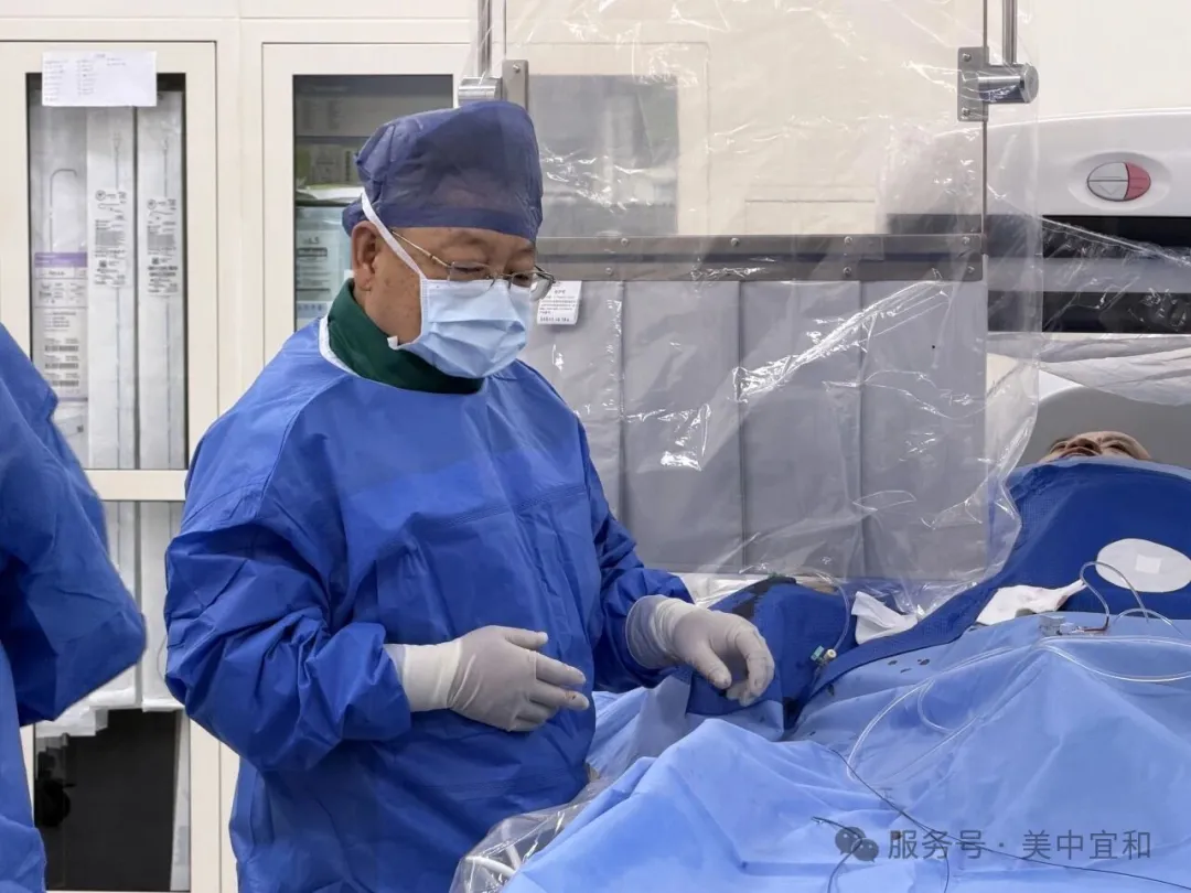
At 9 p.m. the ambulance reached AMCARE Beijing. The emergency team bypassed the waiting room and took Xiao Ai directly to the catheterization laboratory.
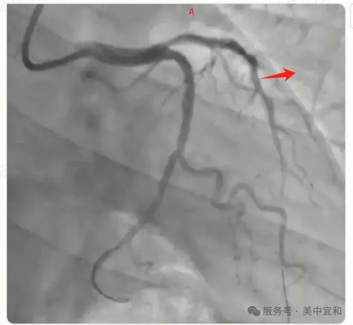
Coronary angiography confirmed the team's fear: the left anterior descending artery was completely occluded. The LAD supplies the anterior wall of the heart; its sudden closure often means extensive myocardial necrosis and a high risk of sudden death. On the screen, the vessel ended abruptly — a river cut in half.
Pre-PCI Angiogram
The left anterior descending artery showed abrupt cutoff, consistent with thrombotic occlusion.
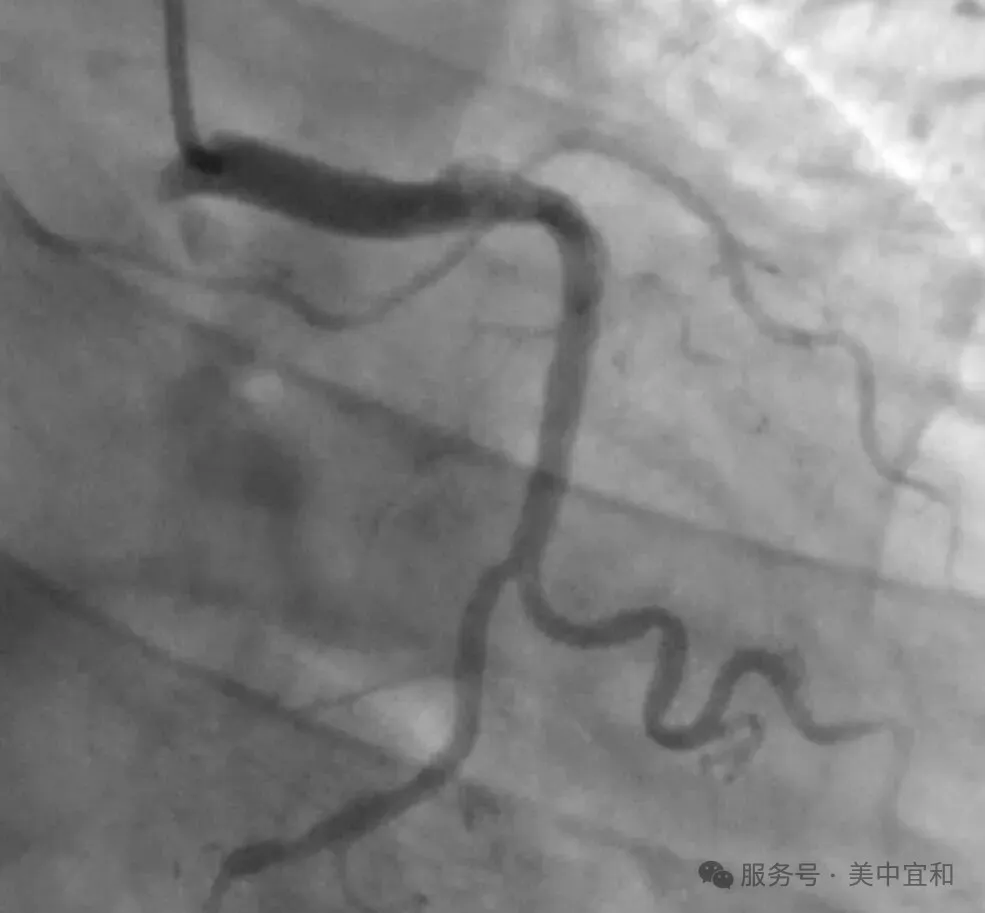
Post-PCI Angiogram
After thrombus aspiration and balloon dilation, contrast refilled the vessel with TIMI grade-3 flow.
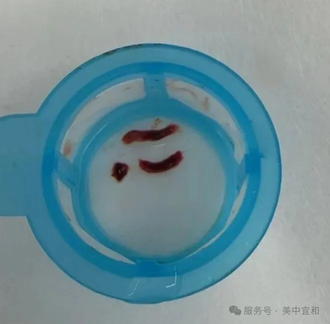
The interventional team moved without hesitation. A suction catheter was advanced, and thrombus was repeatedly aspirated. Then a balloon was inflated to reopen the narrowed segment. As contrast flowed back into the LAD, the vessel filled again: TIMI grade-3 flow, meaning normal myocardial perfusion had been restored.
The Unexpected Decision: No Stent
In routine acute-MI PCI, balloon dilation is usually followed by stent implantation to scaffold the artery and reduce reocclusion. This time, the team stopped after the balloon.
The cardiology director explained the reasoning. First, stenting would have demanded prolonged, intensive dual antiplatelet therapy plus anticoagulation. With Xiao Ai's abdominal wounds barely a day old, stronger antithrombotic drugs could trigger catastrophic postoperative bleeding and recreate the very dilemma the team was trying to escape.
Second, the patient was young, and the underlying mechanism was thromboembolism rather than atherosclerotic stenosis. Once the thrombus was removed and flow restored, the immediate threat was gone. A stent would remain in her artery for life, carrying future risks of in-stent restenosis, in-stent thrombosis, and more complex future procedures. For this specific cause, stenting added long-term risk without clear long-term benefit.
Outcome and Reflection
After the procedure, Xiao Ai was transferred to the ICU for close monitoring. Her cardiac function recovered gradually, and she did not develop serious postoperative bleeding. By the time of discharge, she had passed the acute crisis.
This case illustrates something important beyond the technical success of PCI. When no guideline fits perfectly, clinical decisions must center on the individual: her age, her fresh surgical wounds, her thrombotic mechanism, and her long-term prognosis. MDT collaboration broke down specialty silos and let the team weigh benefit against risk in real time.
"Restoring blood flow early is the key in acute MI. Once TIMI-3 flow is back, the heart is protected. Every subsequent decision — including whether to place a stent — has to be judged by what this specific patient can safely tolerate."
— AMCARE Beijing Cardiology Team

Advances in medicine are not only about better devices. They are also about the discipline to put the patient at the center of complex decisions and to find a safer path when every option carries danger.
Need Urgent Cardiac or International Patient Support?
AMCARE Beijing's emergency and international patient teams coordinate rapid triage, MDT consultation, and post-procedure care for complex cardiac and surgical cases.